Ophthalmology
Ophthalmology
The Ophthalmology Service at the Shreiber School of Veterinary Medicine
What is a Veterinary Ophthalmologist?
A Diplomate of the American College of Veterinary Ophthalmology (DACVO®) requires an extensive amount of education and training. Veterinary ophthalmologists are required to hold a bachelor’s degree, graduate from an accredited four-year veterinary school, complete a rotating internship, and a specialized residency program in ophthalmology. Many veterinary ophthalmologists also complete specialized ophthalmology internships, fellowships, obtain master’s degrees or PhDs during their residency programs, and must meet publication requirements in order to graduate. Veterinary ophthalmologists spend 6 or more years training in the diagnosis and treatment of eye disorders to be eligible to pass a multi-day, multi-part written and practical examination. Once a veterinarian is granted DACVO® status, they must receive regular continuing education in the field of veterinary ophthalmology to achieve Maintenance of Certification. Your board-certified veterinary ophthalmologist has often completed 10+ years of schooling post-college to practice their discipline. This establishes the highest standards of patient care in veterinary ophthalmology.
Book Your Appointment
Call 856-256-6000 (between 8am-8:30pm)
Request an Appointment by Online Form
Request an Appointment by Email
Specialized Care for Animal Eyes at Rowan University
- A board-certified veterinary ophthalmologist at the Shreiber School of Veterinary Medicine of Rowan University can treat any eye with any eye problem. These include dogs, cats, horses, companion small mammals, companions reptiles, companions birds, companion amphibians, sheep, pigs, cows, other livestock, and other companion animals (on a case-by-case basis).
- PLEASE NOTE: We do not accept venomous reptiles, crocodilians, or Old-World primates. We also do not accept native wildlife species, including feral wildlife that pose a rabies risk.
- When eye conditions are found and treated early, this can lessen pain and save vision.
- The ophthalmology team will communicate directly with you and your primary care veterinarian(s) to make sure that your animal receives the best care possible.
What to Expect at Your Visit
- We want your appointment with our veterinary ophthalmology team to be a great experience from the moment you enter our clinic!
- Prior to your appointment, you will receive a brief Patient Visit Questionnaire.
- When you arrive, you will be welcomed by one of our Client Service Representatives who will ask you to complete any necessary intake forms.
- Please have any primary care veterinarians and veterinary specialists that have ever examined your pet fax or email your pet’s medical records, labwork, and surgical records with biopsy results to the Veterinary Teaching Hospital at Rowan University prior to your visit.
- If your pet is covered by pet insurance, you may bring a claim form with you to your appointment.
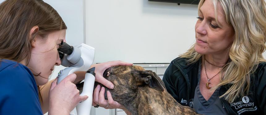
The Eye Exam
- Eye examinations with a board-certified ophthalmologist use specialized equipment. These provide a thorough and microscopic examination of your pet’s eye to diagnose conditions rapidly and accurately, or confirm a previous diagnosis.
- Our examination equipment is digital, and connected to teaching screens in the Ophthalmology Suite within the protected area of the hospital. This means that some of your pet’s exam will be performed away from you.
- Your pet will have baseline diagnostic testing performed at each examination, including a Schirmer tear test, fluorescein stain, and intraocular pressure test. Your pet’s pupils may also be dilated. All tests are performed at the discretion of the ophthalmologist.
- We may repeat some diagnostic testing that was already performed by your primary care veterinarian because it can change over time, with the use of medication, or be interpreted differently with the use of specialized equipment.
- Photographs of the eye are stored in the medical record as part of the exam in order to monitor response to treatment over time (and to compare with photos you may send us from home).
- The result of every diagnostic examination will be shared with you and your primary care veterinarian, and your pet will never undergo a diagnostic examination that is not necessary for making a diagnosis.
Treatments and Surgery
- Once your pet has been examined, your ophthalmologist will discuss all treatment options with you. Some may require medications, and some may require surgery. We will work together to determine what plan is best for your pet and your family.
- Treatments plans that include estimated cost will always be provided to you.
After Your Appointment
- Once your visit is completed, you will be provided with a copy of your pet’s medical records, describing their diagnosis, medication instructions, treatment options, and next recommended recheck examination. These can be printed, emailed to you, or both.
- Our ophthalmologists will send the same copy of your pet’s medical records to your primary care veterinarian’s office, and will also contact your veterinarian personally. It is our mission to make sure that your pet receives the most comprehensive care possible.
Some of the Conditions We Treat
Orbital
- Retrobulbar Abscess, Cellulitis, Tumor, Foreign Body, Cyst
- Nasolacrimal Duct Inflammation, Infection, Stricture, Foreign Body, Cyst
Eyelid and Periocular
- Eyelid Tumors, Cysts
- Blepharitis
- Entropion
- Ectropion
- Distichiasis/Trichiasis
- Ectopic Cilia
- Eyelid Agenesis
- Prolapsed Gland of the Nictitating Membrane (“Cherry Eye”)
- Scrolled Third Eyelid Cartilage
- Dry Eye (KCS) and Evaporative Dry Eye Disease (EDED)
Cornea and Sclera
- Corneal Ulceration
- Corneal Infection
- Corneal Inflammation, Scarring, Pigmentation
- Pannus and Chronic Superficial Keratitis
- Dermoid
- Corneal Perforation, Descemetocele
- Episcleritis, Episclerokeratitis, Scleritis
- Epibulbar Melanoma
Intraocular Disease
- Cataracts
- Lens Luxation or Subluxation
- Iris Freckle, Nevus, Tumor, Melanosis, Melanoma
- Uveitis
- Pigmentary Uveitis, Golden Retriever Uveitis
- Glaucoma
Retina
- Progressive Retinal Atrophy (PRA)
- Retinal Degeneration
- Sudden Acquired Retinal Degeneration (SARDS)
- Retinal Detachment
Some of the Services We Offer
- Slit Lamp Biomicroscopy
- Indirect Ophthalmoscopy
- Tonometry
- Nasolacrimal Duct Irrigation
- Chemical Cyst Ablation*
- Cidofovir Ciliary Body Ablation*
- Enucleation (with or without orbital conformer)
- Intrascleral Prosthesis Placement (ISP)
- Eyelid Mass Debulking with adjunctive Electrocautery and Cryotherapy*
- Periocular Mass Excision with adjunctive Electrocautery*
- Eyelid Filler for Entropion*
- 3-D surgical microscopy
- Surgical microscopy
- Cryomatic cryotherapy
- Keratectomy
- Conjunctival Graft
- Corneoconjunctival Transposition
- Corneal Graft
- Iris Mass Excision/Biopsy
- Diode Laser Therapy for Iris Pigmentation
- Laser Transscleral Cyclophotocoagulation Therapy for Glaucoma
- Gonioimplantation for Glaucoma
- Subconjunctival Implants for Glaucoma*
- Subconjunctival Implants for Pannus and Dry Eye*
- Cataract Surgery with Artificial Lens Implantation
- Specialized Ophthalmic Tissue Biopsy
*commonly offered using oral sedation and/or injectable sedation with topical analgesia (numbing) – no general anesthesia
Current Ophthalmology Clinical Trials
- A New Treatment for Dry Eye in conjunction with Merck Animal Health*
*Must meet eligibility criteria for enrollment; eligible for covered costs of care.
Laura Mancuso, VMD, DACVO®
Clinical Assistant Professor, Ophthalmology
Dr. Laura Mancuso is a clinical assistant professor and board-certified veterinary ophthalmologist at Shreiber School of Veterinary Medicine. She received a Bachelor of Science (Biology) from Virginia Tech in 2011 and graduated from University of Pennsylvania, School of Veterinary Medicine in 2015. After a small animal rotating internship at University of Tennessee, she completed a specialty internship and residency in veterinary ophthalmology at Animal Eye Care of Virginia Beach. Throughout her professional career, she has found the most achievement and reward in mentoring colleagues – students, technicians, interns, and residents – to share her enthusiasm for veterinary ophthalmology.
Read full bio and list of publications on the Clinical Sciences page
Christine Jones, LVT, VTS (Ophthalmology)
Veterinary Technician, Specialty Care
Christine graduated with an Associate of Science degree in Veterinary Technology from Manor College. She was accepted to sit for the first ever VTS (Ophthalmology) exam and passed. Now, she is a Veterinary Technician at the Veterinary Teaching Hospital at New Jersey’s first Veterinary School, the Shreiber School of Veterinary Medicine of Rowan University.
Christine has a special interest in Client Education, which means helping the client with the treatment of their pet as well as helping them understand why the treatment plan is necessary. She also is very interested in incorporating Pain Management and Spectrum of Care alternatives for her patients to keep them most comfortable and/or prioritize their overall health.
Kristen Morris-Green, BS, LVT
Veterinary Technician, Specialty Care
Amy Waters, CVT
Veterinary Technician, Primary and Specialty Care